Interpretation
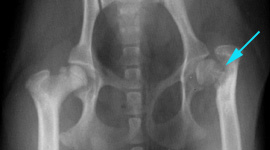
Radiographic Findings:
The right femoral head is irregularly radiolucent at the cranial margin. Both coxofemoral joint spaces are wider than normal. There is a chronic fracture of the left femoral head. There is sclerosis of the subtrochanteric region and proximal portion of the femoral diaphysis. There is reduction in the thickness of soft tissues of the left thigh region.
Radiographic Impressions:
Findings consistent with chronic left femoral neck fracture (see below) with disuse atrophy of the left hind leg. Changes consistent with bilateral Legg-Calve-Perthes disease.
Post Operative:
Left sided femoral head and neck ostectomy and stifle surgery for repair of a left sided medial patellar luxation.
J.M. Goggin, DVM, DACVR