Lameness Locator
Jarrod Younkin, DVM, Equine Intern, Veterinary Health Center at Kansas State University
|
See more on YouTube! |
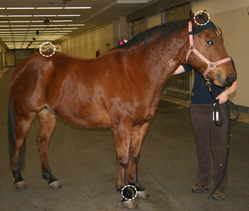 |
| Figure 1: Example of a horse instrumented with the Lameness Locator system. Accelerometers have been placed on the top of the head and croup. The gyroscope has been placed on the pastern of the right front limb. The 3 devices are highlighted by white dash-lined circles. |
 |
|
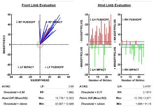
Figure 2: Example of the Lameness Locator read out on a horse during a lameness work-up. The chart figures go through different detections of lameness for front leg evaluations (left side line-graph) and hind leg evaluations (right side bar-graphs). The veterinarian will go through the lameness with the owner and explain Lameness Locator findings. |
Equine lameness is a common reason that horse owners and trainers seek out veterinary care. When a horse is examined for musculoskeletal disease a complete lameness exam is performed. In general terms, lameness is subjectively evaluated by the examining veterinarian, which allows for the determination of disease severity by being placed into an AAEP lameness grading scale from 1-5. Grade 1 is the most mild, while a horse suffering from grade 5 lameness will be unable bear weight (most severe). Although this system of assessment is useful, it is not a perfect method of evaluation. Therefore, alternate methods of lameness evaluation have been investigated in an effort to apply an objective measurement to patient assessment. Among the methods that have been studied, a camera-based kinetic evaluation of the horse on a treadmill has been tested using a stationary force plate. This kinematic system paved the groundwork for the current system which utilizes a wireless inertial sensor system that is currently being implemented at the VHC.
The lameness locator is an inertial sensor system that utilizes two accelerometers, one placed on the top of the croup and another on the poll of the head and one gyroscope placed on the right forelimb pastern (Figure 1). These sensors accurately detect and quantify forelimb and hind limb lameness when the horse is trotting in a straight line as well as lunging in a circle. The inertial sensor data is transmitted to a tablet computer so that kinematic algorithms can be performed on the collected sensor data. The detection and quantification of the horse’s lameness is reported as a ratio of vertical motion and height differences of the head and pelvis. The results are displayed as numbers and x and y graphs where right and left front or hind limb lameness can be measured (Figure 2).
Difficult lameness exams such as demonstrated in horses with a head nod (front end lameness) and an asymmetric hip excursion (hind end lameness) can be challenging to diagnose; therefore, the lameness locator provides objective measures that can aid in the determination of the source(s) of lameness. One reason that horses may appear to be lame in front and behind may actually be a result of a primary lameness with a compensatory response in the other area. For instance, in some cases the primary lameness is in the front half of body and yet compensatory movements are present in the back half (hind end) which result in the appearance of lameness in both places. This phenomena is referred to as the “Law of Sides”. Although this can be a challenging scenario from a clinical and diagnostic standpoint, the lameness locator provides the clinician with the ability to decrease confusion regarding the primary lameness and more accurately determine the primary source of pain.
Overall, the objective measurements of equine lameness that are acquired by the lameness locator can aid equine clinicians with the diagnosis of lameness that range in severity from mild to severe. The lameness locator is not a substitute for the equine veterinarian evaluation of lameness, it is rather an aid for equine veterinarians and owners to detect and evaluate challenging cases of equine lameness.
- USDA. National economic cost of equine lameness, colic, and equine protazoal myeloencephalitis in the United States. Information sheet No. N348.1001. Fort Collins, Colo: USDA APHIS Veterinary Services National Health Monitoring System, 2001.
- Keegan KG. The Lameness Locator (wireless inertial sensors for detection of lameness in horses). WVOC. 215-217 2010
- Keegan KG et. al. Assessment of repeatability of a wireless, inertial sensor-based lameness evaluation system for horses. AJVR Vol 72, No. 9, 2011
- Keegan KG et. al. Comparison of an inertial sensor system of lameness quantification with subjective lameness evaluation. EVJ 44 2012